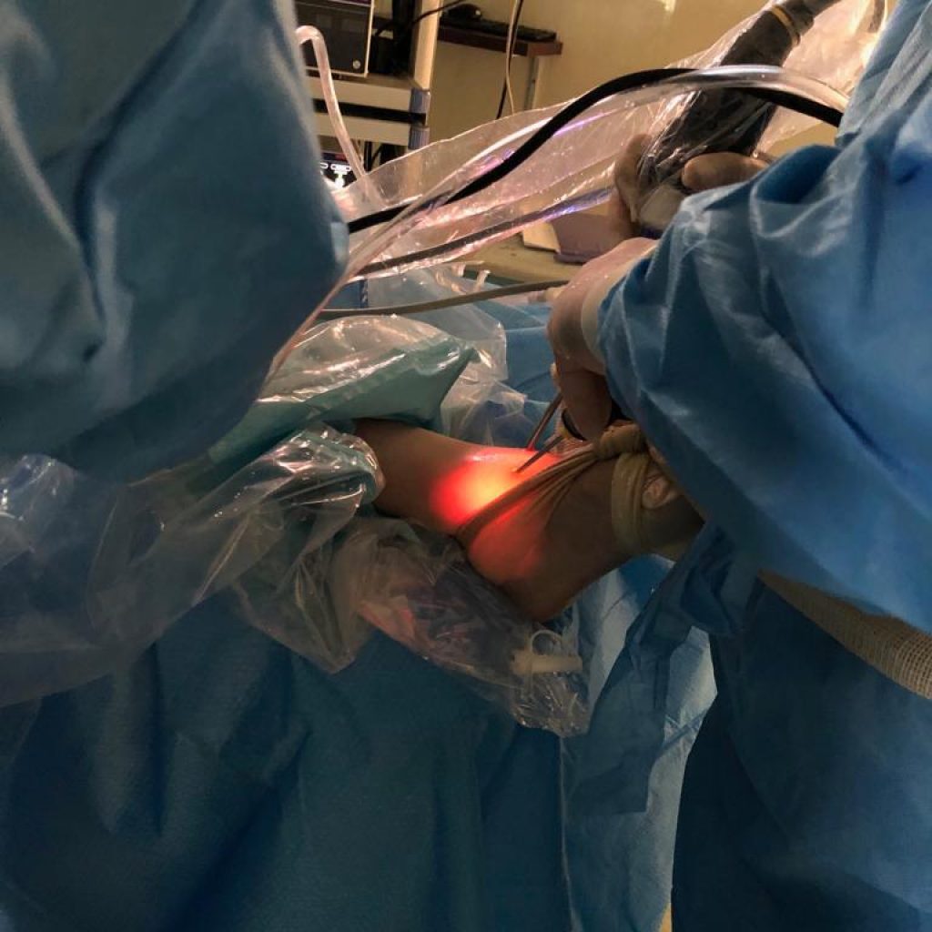
Foot and Ankle Disorders

Arthroscopic & open view of the TFAL. Rapture, RT. Ankle

TFAL. Reconstruction using two ancores

Forefoot Hematoma

Open Extirpation

Situation Before Wound Closure

X-ray image ap. & lat. view mid foot trauma with complete into medial dislocation of the Os Naviculare LT.

Intra operative situs from medial approach

Positioning of the distractor

Exposed and removed Navicular bone

Ap. C-Arm Radiography shows the empty place of the bone and exact Position of the K.W.

The mid foot place is ready for the repositioning of the bone

Os Naviculare is back to its anatomical position

X-Ray image ap. Shows also the good position

Fixation of the mid foot with retrograde titanium long screw from trans Metatarsus I. Distal

Additional fixation with titanium mini Plate

Additional fixation with titanium mini Plate

X-ray image ap. Shows the final fixation result

Wound closure

Chairman:

Dr. Gh. Naderi
Orthopaedic Surgeon

E-Mail:
[email protected]

Website:
www.dr-rezanaderi.com
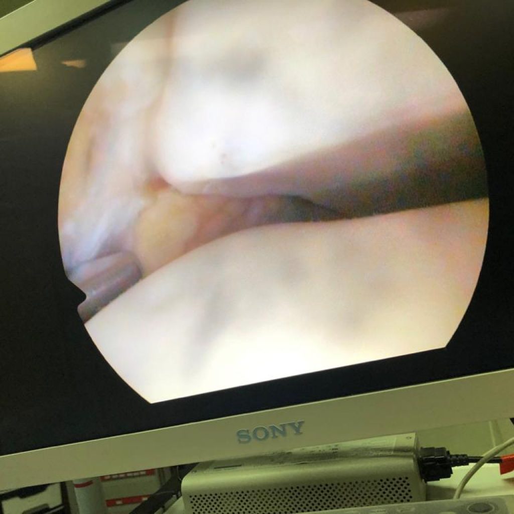
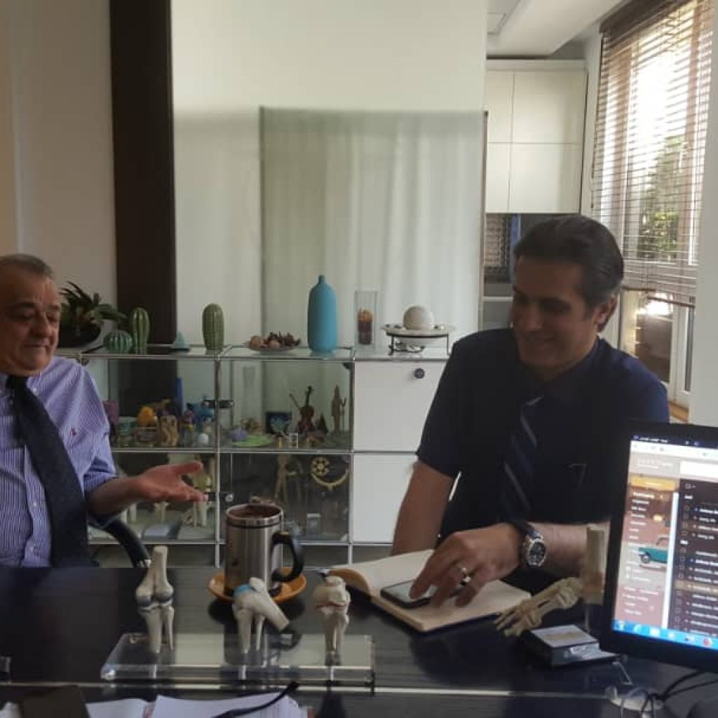
From Left: Prof. Dr. F. Farid, Dr. Gh. Naderi

From Left: Dr.  Gh. Naderi, Prof. Dr. F. Farid

From Left: Dr.  Gh. Naderi, W. Schäfer, Prof. Dr. F. Farid